Researchers from the University of Granada, Spain, and the University of Glasgow, Scotland, have used 3D printing to enable the diagnosis of parasitic infections using smartphones.
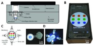
Utilizing a specifically developed Android-based software application, the platform is able to make an automatic and accurate analysis of the Trypanosomatid species using the smartphone’s rear camera. Trypanosomatids are parasites responsible for a number of diseases in humans and livestock. This diagnosis can be achieved on the mobile phone using a 3D printed plastic accessory, which attaches to the smartphone camera and provides controlled illumination and fixed sample positioning.
By developing an accessible and affordable method for diagnosing these diseases, the researchers hope that it can be utilized in remote areas of developing countries that have limited access to resources for such tools. In the research paper published in IEEE Access, the authors state:
“ITS SIMPLICITY AND RELIABLE PERFORMANCE MEAN IT CAN BE USED IN REMOTE, LIMITED-RESOURCE SETTINGS BY RELATIVELY UNSKILLED TECHNICIANS/NURSES, WHERE DIAGNOSTIC LABORATORIES ARE SPARSELY DISTRIBUTED. THE RESULTS CAN HOWEVER BE SENT EASILY VIA THE SMARTPHONE TO MEDICAL EXPERTS AS WELL AS GOVERNMENT HEALTH AGENCIES.”
The researchers explain that smartphones are powerful, accessible and easy-to-use devices that can “significantly enhance medical and animal health diagnostic abilities, particularly in settings with limited resources.” Their ability to quickly transmit data can also be leveraged for fast transfer of analysis results to health care personnel, enabling the diagnosis of diseases in areas without the infrastructure to support fast analysis.
The research team also explains that these smartphone cameras have the potential to be used for colorimetric responsive assays, which are used to measure any test substance that is itself colored or can be reacted to produce a color. Colorimetric responsive assays can be leveraged to detect disease biomarkers, and smartphones have been utilized in such applications for the detection of tuberculosis and HIV, according to the paper.
However, the paper explains that “various factors can affect the reliability of image processing and as a reliable diagnostic tool.” As such, the researchers propose an external attachment be used to ensure performance specifications are met, as equipment-free methods can suffer from poor repeatability as well as standardization problems due to reflections and sensor positioning. Therefore, the researchers opted to 3D print a plastic accessory as a low-cost solution for providing controlled illumination and fixed sample and cell phone holding.
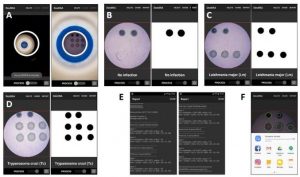
As well as the 3D printed accessory, the research team also developed an Android-based software application for the smartphone in order to test its capabilities of analyzing millimetric colorimetric arrays. The application is able to recognize and quantify the color coordinates of each spot in the array pattern through the phone’s camera.
Increasing access to medical diagnostic testing
Focusing on parasitic diseases of the family Trypanosomatidae, the researchers put their smartphone diagnostic platform to the test using real DNA. These parasites affect both humans and livestock, and they can lead to significant health and economic consequences for many countries. Using the camera of a smartphone provides various benefits for detecting such diseases beyond accessibility, including a reduction of human errors in pattern recognition as a result of untrained staff. It also enables the creation of a permanent record to be shared with health experts.
From the tests, the researchers concluded that with the 3D printed accessory and Android application, the smartphone was able to accurately detect Trypanosomatid diseases in real DNA. Their platform was tested repeatedly to evaluate its size detection, edge blurriness, and color detection capabilities, validating its use for the analysis of colorimetric spot arrays. The 3D printed accessory enables the camera to detect spots with diameters from 300 µm with a 15 percent tolerance.
Concluding the paper, the researchers wrote: “This system has been tested for detection of parasitic diseases of the family Trypanosomiases, which are responsible for devastating diseases in humans, dogs as well as livestock. In this context, the smartphone-based platform that has been developed analyses the image and converts the panel of spots into a Trypanosomatid species,”.
“GIVEN THE IMPORTANCE OF SUCH ASSAYS IN DEVELOPING COUNTRIES, THE MOBILE PHONE IMAGER APPLICATION HAS BEEN DESIGNED TO BE ESPECIALLY USER-FRIENDLY FOR ITS USE BY UNTRAINED PERSONNEL. RECORDED RESULTS CAN BE IMMEDIATELY TRANSMITTED TO REFERENCE CLINICIANS FROM REMOTE LOCATIONS FOR ADVICE AND TREATMENT DECISIONS.”
This is not the first instance where 3D printing has provided a means for researchers to develop specific, low-cost accessories for mobile phones that transform the devices into useful, accessible medical tools. For example, RMIT University scientists developed a 3D printed “clip-on” filter that can turn smartphone cameras into a powerful microscope. Capable of viewing specimens as small as 1/200th of a millimeter, the device can potentially prove useful as a point of care diagnostic tool or research device, for remote healthcare clinics and field research groups.
Outside of smartphones, 3D printing has also been leveraged to build a standalone high-resolution digital holographic microscopy (DHM) microscope. Seeking to create a portable, powerful and cost-effective microscope, U.S. researchers 3D printed the device to enable the diagnosis of diseases like malaria, sickle cell disease, diabetes, and others.
The simplicity and low cost of constructing the instrument, which is made entirely from 3D printed parts and commonly found optical components, could “increase access to low-cost medical diagnostic testing,” according to research team leader Bahram Javidi from the University of Connecticut, which he claims “would be especially beneficial in developing parts of the world where there is limited access to health care and few high-tech diagnostic facilities.”

A) Lateral schematic view and B) photograph of the complete setup composed of the 3D printed accessory, the illuminated centrifuge collection tube, and the smartphone. C) Top schematic view and D) photographs of the centrifuge collection tube with the white LEDs.
App screenshots.
Image acquisition and processing workflow in the smartphone application.
ENLACES DE INTERES
Referencia bibliográfica
“Smartphone-based diagnosis of parasitic infections with colorimetric assays in centrifuge tubes” is written by Pablo Escobedo, Miguel M. Erenas, Antonio Martínez Olmos, Miguel A. Carvajal, Mavys Tabraue Chávez, Angélica Luque González, Juan J. Díaz-mochón, Salvatore Pernagallo, Luis Fermín Capitán-vallvey, and Alberto J. Palma.


